ASK US A QUESTION
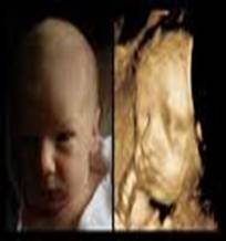
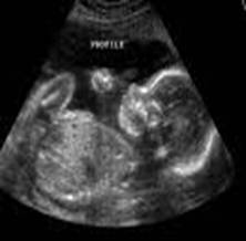
MEDICAL SONOGRAPHY (ULTRASONOGRAPHY) is an ultrasound-based diagnostic medical imaging technique used to visualize muscles, tendons, and many internal organs, to capture their size, structure and any pathological lesions with real time tomographic images. Ultrasound has been used by radiologists and sonographers to image the human body for at least 50 years and has become one of the most widely used diagnostic tools in modern medicine. Ultrasound is used to visualize fetuses during routine and emergency prenatal care. Such diagnostic applications used during pregnancy are referred to as obstetric sonography.
Obstetric ultrasound can be used to identify many conditions that would be harmful to the mother and the baby. Many health care professionals consider the risk of leaving these conditions undiagnosed to be much greater than the very small risk, if any, associated with undergoing an ultrasound scan. According to Cochrane Review, routine ultrasound in early pregnancy (less than 24 weeks) appears to enable better gestational age assessment, earlier detection of multiple pregnancies and earlier detection of clinically unsuspected fetal malformation at a time when termination of pregnancy is possible.
Obstetric ultrasound is primarily used to:
- Date the pregnancy
- Confirm fetal viability
- Determine location of fetus, intrauterine vs. ectopic
- Check the location of the placenta in relation to the cervix
- Check for the number of fetuses
- Check for major physical abnormalities.
- Assess fetal growth (for evidence of intrauterine growth restriction (IUGR))
- Check for fetal movement and heartbeat.
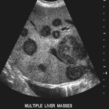
As currently applied in the medical field, properly performed ultrasound poses no known risks to the patient. Sonography is generally described as a "safe test" because it does not use mutagenic ionizing radiation, which can pose hazards such as chromosome breakage and cancer development. In 2008, the AIUM published a 130-page report titled "American Institute of Ultrasound in Medicine Consensus Report on Potential Bioeffects of Diagnostic Ultrasound" stating that long term effects due to ultrasound exposure at diagnostic intensity is still unknown. It should be noted that obstetrics is not the only use of ultrasound. Soft tissue imaging of many other parts of the body is conducted with ultrasound. Other scans routinely conducted are cardiac, renal, liver and gallbladder. Other common applications include musculo-skeletal imaging of muscles, ligaments and tendons, ophthalmic ultrasound (eye) scans and superficial structures such as testicle, thyroid, salivary glands and lymph nodes. Because of the real time nature of ultrasound, it is often used to guide interventional procedures such as fine needle aspiration FNA or biopsy of masses for cytology or histology testing in the breast, thyroid, liver, kidney, lymph nodes, muscles and joints. According to the European Committee of Medical Ultrasound Safety (ECMUS) "Ultrasonic examinations should only be performed by competent personnel who are trained and updated in safety matters.
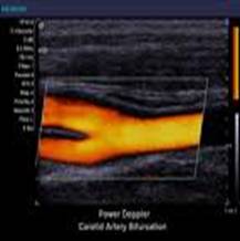
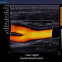
Medicare Ultrasound scanners have different Doppler-techniques to visualize arteries and veins. The most common is colour Doppler or power doppler, but also other techniques like b-flow are used to show blood flow in an organ. By using pulsed wave Doppler or continuous wave doppler blood flow velocities can be calculated. Ultrasound is also increasingly being used in trauma and first aid cases, with emergency ultrasound becoming a staple of most EMT response teams.
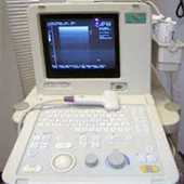
Medicare Investigations (P) Ltd, India, owned centers are well equipped with “State of art” Diagnostic Ultrasound and Color Doppler Machines including ACUSON ASPEN, PHILLIPS, TOSHIBA and HITACHI Scanners at its various centers in India. Best of all that the Radiologists behind these Scanners are well trained and long time experienced with institutional back up and to perform all kind of investigations up to tertiary levels for the benefit of the public since more than 17 years.
